

Technology
Decay Risk Assessment
The emerging biofilm science is changing how we look at decay as a disease model. The new technologies in our office better assess, detect and diagnose signs of disease presence, progression and activity levels.
Typical Operatories at Johansen Dentistry
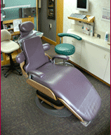 Typical Opertory Room |
Panorex X-Ray
The Panorex X-ray is a single picture of all your teeth and surrounding bones. Sometimes referred to as a single FMX, or full mouth X-ray, the Panorex provides a two-dimensional panoramic view of your mouth. The resulting X-ray includes more than just a couple of teeth at a time -- and is an excellent alternative to the tiny pictures your dentist has to piece together to see a complete set of your teeth.
The Panorex X-ray also exposes parts of your jaw that can't be seen with traditional dental X-rays. With a Panorex single FMX, your dentist can view:
- Your entire upper and lower jawbone
- Your temporomandibular joints (TMJ), or jaw joints
- The nasal sinuses and their surrounding bone
- The mandibular nerve, which provides sensation to the teeth and gums of the lower jaw
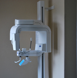 Panorex Device |
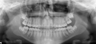 Panorex X-Ray Example |
Digital X-Ray
In addition to much
lower radiation exposure, there is no film to develop with
digital X-Rays. Instead, images are captured instantly in
our computer system. The images can then be moved around,
enlarged and manipulated. This allows Dr. Johansen and the patient to evaluate the conditions of the
oral cavity for proper diagnosis. Patients appreciate being
able to instantly view their X-Rays on the wall-mounted
monitors.
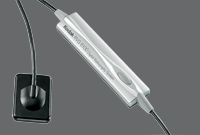 Kodak Digital X-Ray Receiver |
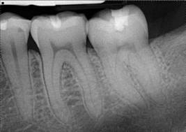 Digital X-Ray Example |
IntraOral Camera
Our IntraOral Camera technology allows Dr. Johansen to take full 3-Dimensional photos of your teeth and
gums including lesions, cracks, and calculus buildup.
 IntraOral Camera Device |
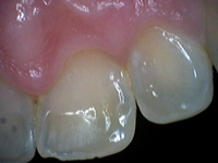
 IntraOral Image Examples |